Classic playlist (2014-2020)
2020
- Fan, Audrey P., Khalil AA, Fiebach JB, Zaharchuk G, Villringer A, Villringer K, and Gauthier CJ. Journal of Cerebral Blood Flow & Metabolism 40, no. 3 (2020): 539-551.
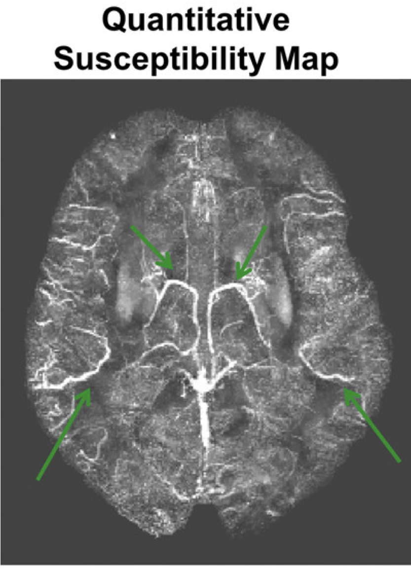
Elevated brain oxygen extraction fraction measured by MRI susceptibility relates to perfusion status in acute ischemic stroke
We showed that MRI can non-invasively quantify the brain's oxygen extraction fraction (OEF) in stroke patients. Brain OEF relates to the blood flow status in these patients, and is sensitive to OEF changes over time after treatment.
- Ishii Y, Thamm T, Guo J, Khalighi MM, Wardak M, Holley D, Gandhi H, Park JH, Shen B, Steinberg GK, Chin FT, Zaharchuk G, Fan, Audrey P. "Simultaneous phase‐contrast MRI and PET for noninvasive quantification of cerebral blood flow and reactivity in healthy subjects and patients with cerebrovascular disease." Journal of Magnetic Resonance Imaging 51, no. 1 (2020): 183-194.
- Chen DYT, Ishii Y, Fan, Audrey P., Guo J, Zhao MY, Steinberg GK, Zaharchuk G. "Predicting PET cerebrovascular reserve with deep learning by using baseline MRI: a pilot investigation of a drug-free brain stress test." Radiology 296, no. 3 (2020): 627-637.
- Fan, Audrey P., An H, Moradi F, Rosenberg J, Ishii Y, Nariai T, Okazawa H, Zaharchuk G. "Quantification of brain oxygen extraction and metabolism with [15O]-gas PET: A technical review in the era of PET/MRI." Neuroimage (2020): 117136.
- Mormino EC, Toueg TN, Azevedo C, Castillo JB, Guo W, Nadiadwala A, Corso NK, Hall JN, Fan, Audrey P., Trelle AN, Harrison MB, Hunt MP, Sha SJ, Deutsch G, James ML, Fredericks CA, Koran ME, Zeineh MM, Poston K, Greicius MD, Khalighi MM, Davidzon GA, Shen B, Zaharchuk G, Wagner AD, Chin FT. European Journal of Nuclear Medicine and Molecular Imaging (2020): 1-12.
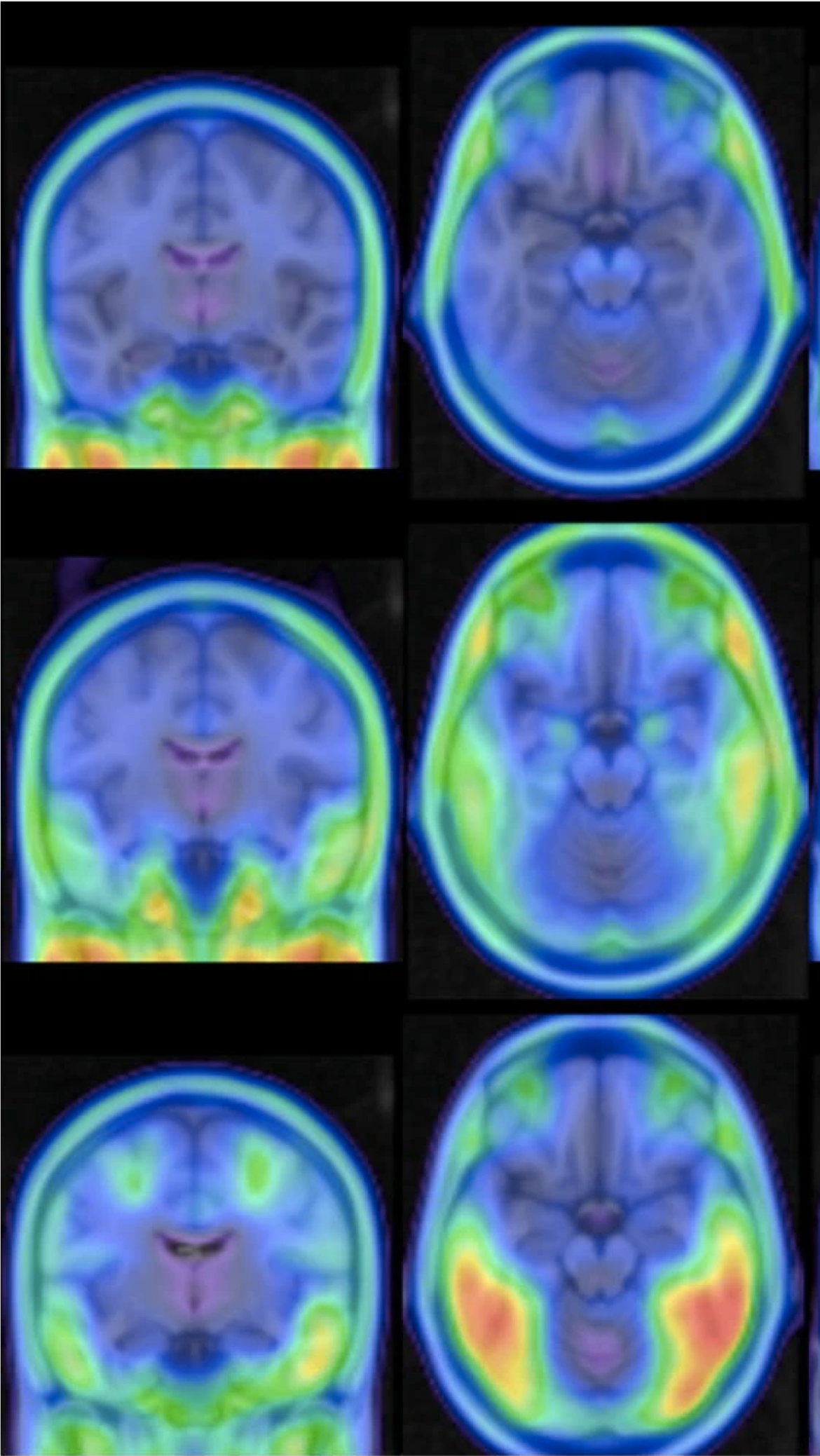
Tau PET imaging with 18 F-PI-2620 in aging and neurodegenerative diseases
We used a second-generation PET tracer, 18F-PI-2620, to non-invasively visualize where tau tangles accumulate in the brain. In patients with cognitive impairment, strong differences in the images were seen in the medial temporal lobe and cortical regions known to be impacted in Alzheimer's Disease.
- Guo J, Gong E, Fan, Audrey P., Goubran M, Khalighi MM, Zaharchuk G. "Predicting 15O-water PET cerebral blood flow maps from multi-contrast MRI using a deep convolutional neural network with evaluation of training cohort bias." Journal of Cerebral Blood Flow & Metabolism 40, no. 11 (2020): 2240-2253.
- Carlson ML, DiGiacomo PS, Fan, Audrey P., Goubran M, Khalighi MM, Chao SZ, Vasanawala M, Wintermark M, Mormino EC, Zaharchuk G, James ML, Zeineh MM. "Simultaneous FDG-PET/MRI detects hippocampal subfield metabolic differences in AD/MCI." Scientific reports 10.1 (2020): 1-7.
2019
- Fan, Audrey P., Khalighi MM, Guo J, Ishii Y, Rosenberg J, Wardak M, Park JH, Shen B, Holley D, Gandhi H, Haywood T, Singh P, Steinberg GK, Chin FT, Zaharchuk G. Stroke 50.2 (2019): 373-380.
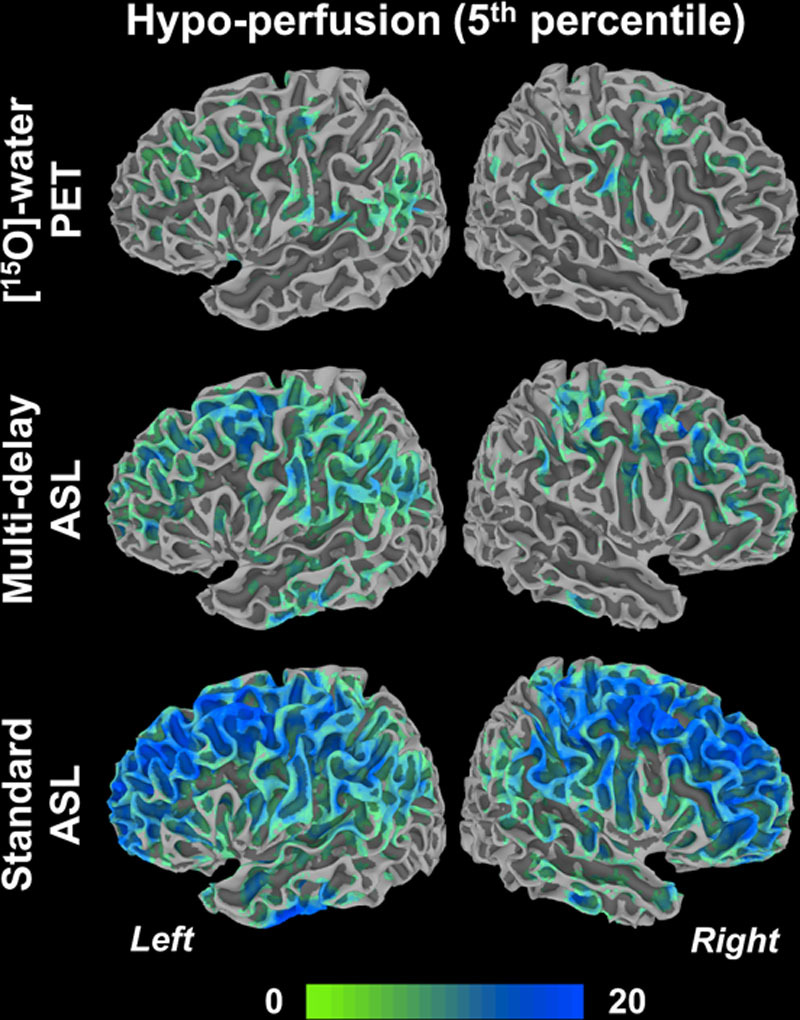
Identifying Hypoperfusion in Moyamoya Disease With Arterial Spin Labeling and an [15O]-Water PET/MRI Normative Database.
Arterial spin labeling (ASL) is an MRI-based imaging method to quantify cerebral blood flow in the brain, including in patients with cerebral artery blockages. We showed ASL can identify poor blood flow in patients, similar to the PET reference standard, if suitable post-label delays are used.
- Gauthier CJ, and Fan, Audrey P. "BOLD signal physiology: models and applications." NeuroImage 187 (2019): 116-127.
- Huck H, Wanner Y, Fan, Audrey P., Jäger AT, Grahl S, Schneider U, Villringer A, Steele CJ, Tardif CL, Bazin PL, Gauthier CJ. "High resolution atlas of the venous brain vasculature from 7 T quantitative susceptibility maps." Brain Structure and Function 224, no. 7 (2019): 2467-2485.
2018
- Kogan F, Fan, Audrey P., Monu U, Iagaru A, Hargreaves BA, Gold GE. "Quantitative imaging of bone–cartilage interactions in ACL-injured patients with PET–MRI." Osteoarthritis and cartilage 26, no. 6 (2018): 790-796.
- Donahue MJ, Achten E, Cogswell PM, Leeuw FE, Derdeyn CP, Dijkhuizen RM, Fan, Audrey P. et al. "Consensus statement on current and emerging methods for the diagnosis and evaluation of cerebrovascular disease." Journal of Cerebral Blood Flow & Metabolism 38, no. 9 (2018): 1391-1417.
- Khalighi MM, Deller TW, Fan, Audrey P., Gulaka PK, Shen B, Singh P, Park JH, Chin FT, Zaharchuk G. Journal of Cerebral Blood Flow & Metabolism 38, no. 1 (2018): 126-135.
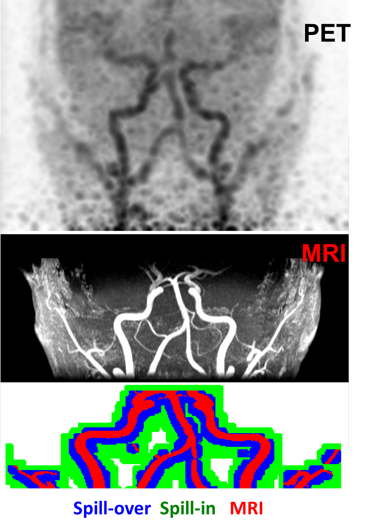
Image-derived input function estimation on a TOF-enabled PET/MR for cerebral blood flow mapping
To quantify blood flow maps from PET, usually arterial blood sampling is needed to understand radiotracer input, but this can be painful, invasive, and challenging. We proposed a new method to get the tracer input directly from arteries in PET and MRI images, without the need for invasive sampling.
- Guo Jia, Holdsworth SJ, Fan, Audrey P., Lebel MR, Zun Z, Shankaranarayanan A, Zaharchuk G. "Comparing accuracy and reproducibility of sequential and Hadamard‐encoded multidelay pseudocontinuous arterial spin labeling for measuring cerebral blood flow and arterial transit time in healthy subjects: a simulation and in vivo study." Journal of Magnetic Resonance Imaging 47, no. 4 (2018): 1119-1132.
2017
- Fan, Audrey P., Jia Guo, Mohammad M. Khalighi, Praveen K. Gulaka, Bin Shen, Jun Hyung Park, Harsh Gandhi et al. "Long-delay arterial spin labeling provides more accurate cerebral blood flow measurements in moyamoya patients: a simultaneous positron emission tomography/MRI study." Stroke 48, no. 9 (2017): 2441-2449.
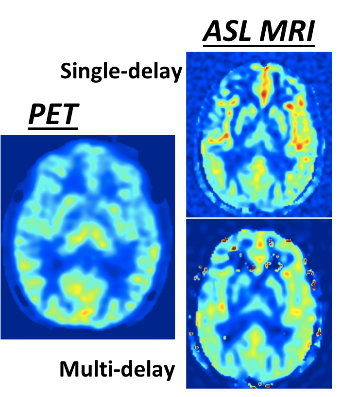
Long-delay arterial spin labeling provides more accurate cerebral blood flow measurements in moyamoya patients: a simultaneous PET/MRI study.
We used simultaneous PET and MRI to validate scans of brain blood flow. We performed PET/MRI scans in challenging Moyamoya cases, who have blocked arteries and very long arterial transit times. Clinically, we recommend arterial spin labeling MRI with long post-label delays and multiple-delay time points for these patients.
- Kogan F, Fan, Audrey P., McWalter EJ, Oei EHG, Quon A, Gold GE. "PET/MRI of metabolic activity in osteoarthritis: a feasibility study." Journal of Magnetic Resonance Imaging 45, no. 6 (2017): 1736-1745.
- Wehrli FW, Fan, Audrey P., Rodgers ZB, Englund EK, Langham MC. "Susceptibility‐based time‐resolved whole‐organ and regional tissue oximetry." NMR in biomedicine 30, no. 4 (2017): e3495.
- McDaniel P, Bilgic B, Fan, Audrey P., Stout JN, Adalsteinsson E. "Mitigation of partial volume effects in susceptibility‐based oxygenation measurements by joint utilization of magnitude and phase (JUMP)." Magnetic resonance in medicine 77, no. 4 (2017): 1713-1727.
- Ward PGD, Fan, Audrey P., Raniga P, Barnes DG, Dowe DL, Ng ACL, Egan GF. "Improved quantification of cerebral vein oxygenation using partial volume correction." Frontiers in neuroscience 11 (2017): 89.
- Soman S, Bregni JA, Berkin Bilgic B, Nemec U, Fan, Audrey P., Liu Z, Barry RL, Du J, Main K, Yesavage J, Adamson MM, Moseley M, Wang Y. "Susceptibility-based neuroimaging: standard methods, clinical applications, and future directions." Current radiology reports 5, no. 3 (2017): 11.
2016
- Fan, Audrey P., Hesamoddin Jahanian, Samantha J. Holdsworth, and Greg Zaharchuk. Journal of Cerebral Blood Flow & Metabolism 36, no. 5 (2016): 842-861.
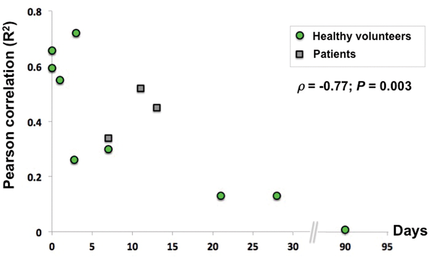
Comparison of cerebral blood flow measurement with [15O]-water positron emission tomography and arterial spin labeling magnetic resonance imaging: a systematic review
We performed a meta-analysis of studies that compared MRI and PET for cerebral blood flow measurements. A major conclusion is that because blood flow varies normally by time-of-day, diet, etc., simultaneous PET/MRI is important to validate these physiological imaging biomarkers.
- Fan, Audrey P., Schäfer A, Huber L, Lampe L, von Smuda S, Möller HE, Villringer A, Gauthier CJ. "Baseline oxygenation in the brain: correlation between respiratory-calibration and susceptibility methods." Neuroimage 125 (2016): 920-931.
- Bazin PL, Plessis V, Fan, Audrey P., Villringer A, Gauthier CJ. "Vessel segmentation from quantitative susceptibility maps for local oxygenation venography." In 2016 IEEE 13th International Symposium on Biomedical Imaging (ISBI), pp. 1135-1138. IEEE, 2016.
2015
- Fan, Audrey P., Evans KC, Stout JN, Rosen BR, Adalsteinsson E. "Regional quantification of cerebral venous oxygenation from MRI susceptibility during hypercapnia." Neuroimage 104 (2015): 146-155.
- Fan, Audrey P., Govindarajan ST, Kinkel RP, Madigan NK, Nielsen AS, Benner T, Tinelli E, Rosen BR, Adalsteinsson E, Mainero C. "Quantitative oxygen extraction fraction from 7-Tesla MRI phase: reproducibility and application in multiple sclerosis." Journal of Cerebral Blood Flow & Metabolism 35, no. 1 (2015): 131-139.
- Bilgic B, Gagoski BA, Cauley SF, Fan, Audrey P., Polimeni JR, Grant PE, Wald LL, Setsompop K. "Wave‐CAIPI for highly accelerated 3D imaging." Magnetic resonance in medicine 73, no. 6 (2015): 2152-2162.
2014 and earlier
- Fan, Audrey P., Bilgic B, Gagnon L, Witzel T, Bhat H, Rosen BR, Adalsteinsson E. "Quantitative oxygenation venography from MRI phase." Magnetic resonance in medicine 72.1 (2014): 149-159.
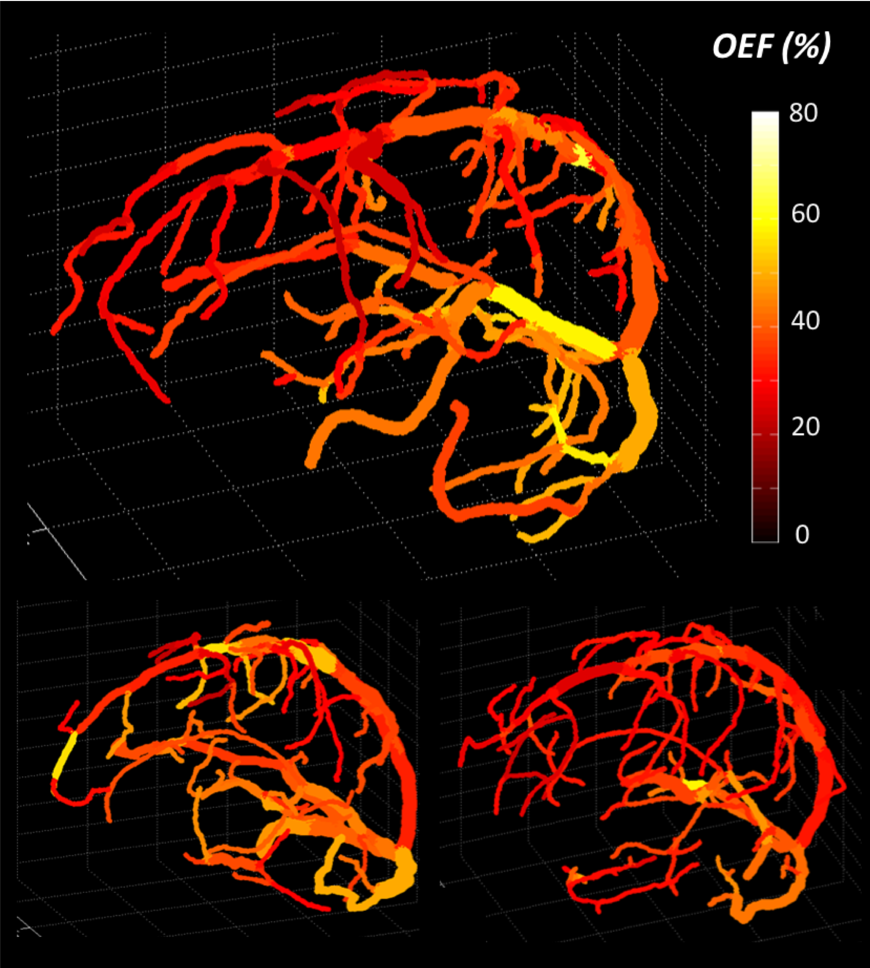
Quantitative oxygenation venography from MRI phase
We developed a novel MRI acquisition and reconstruction approach to map quantitative oxygen extraction fraction (OEF) along cerebral veins. This approach provides non-invasive, local OEF information from the phase image of an accessible gradient echo sequence.
- Bilgic B, Fan, Audrey P., Polimeni JR, Cauley SF, Bianciardi M, Adalsteinsson E, Wald LL, Setsompop K. "Fast quantitative susceptibility mapping with L1‐regularization and automatic parameter selection." Magnetic resonance in medicine 72, no. 5 (2014): 1444-1459.
- Fan, Audrey P., Benner T, Bolar DS, Rosen BR, Adalsteinsson E. "Phase‐based regional oxygen metabolism (PROM) using MRI." Magnetic resonance in medicine 67, no. 3 (2012): 669-678.
